
Audio overview — how insulin sensitivity works, what disrupts it, and what you can do about it
Read the full deep-dive
What Is Insulin Sensitivity & Glucose Regulation?
You used to eat a meal and move on with your day. Now there is a crash two hours later — the brain fog, the irritability, the pull toward something sweet just to get through the afternoon. You are exercising more but seeing less change in your body composition. The weight around your midsection appeared seemingly overnight and refuses to respond to the strategies that used to work.
These experiences are not failures of discipline. They are signals from a biological system that governs how every cell in your body accesses energy — insulin sensitivity and glucose regulation. This interconnected network of hormones, receptors, signaling pathways, and feedback loops determines how efficiently your cells absorb fuel from your bloodstream, how your liver manages blood sugar between meals, and whether your body is primed to store fat or burn it.
Understanding this biology changes the conversation. It moves you from “why isn’t this working?” to “what is actually happening in my body?” — and that shift is where meaningful, lasting progress begins.
How Does It Work?
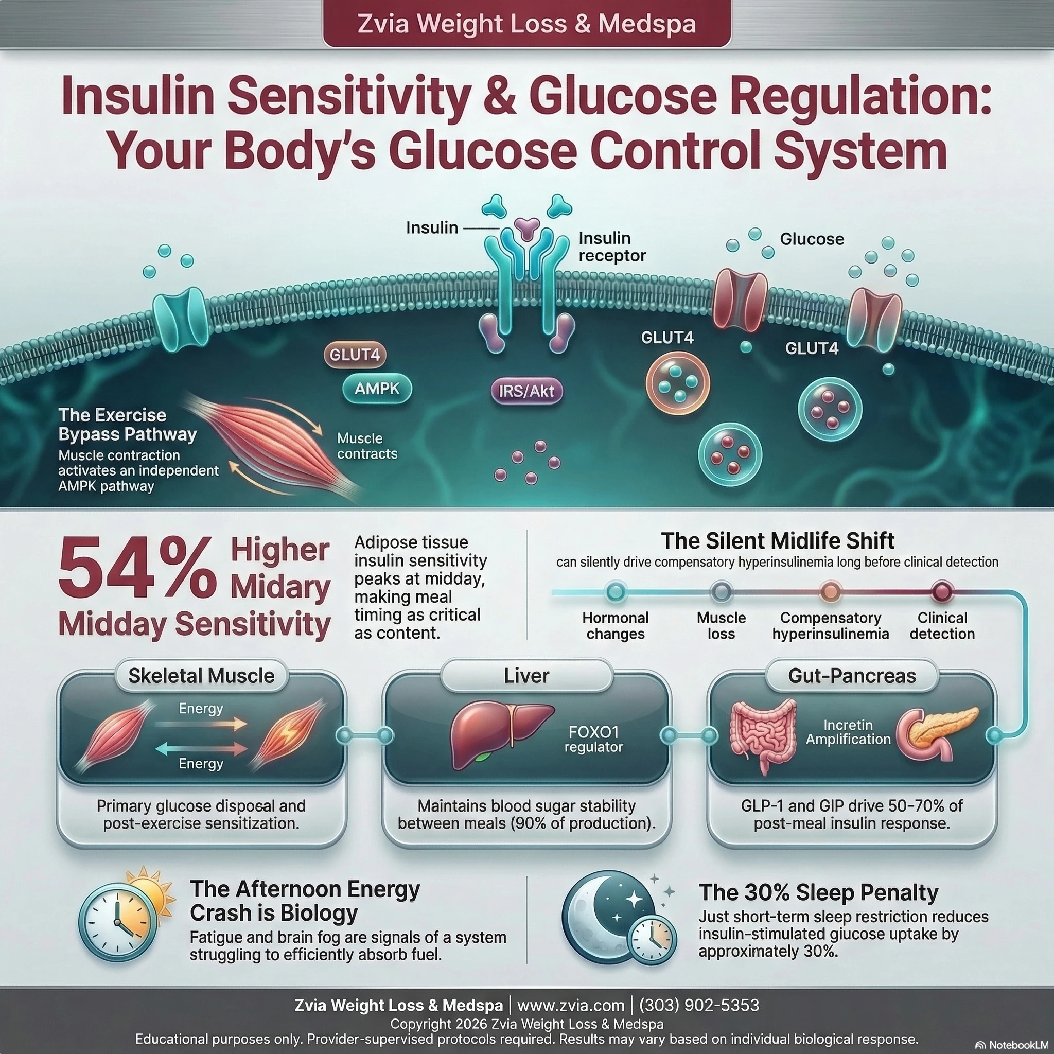
The Insulin Signaling Cascade
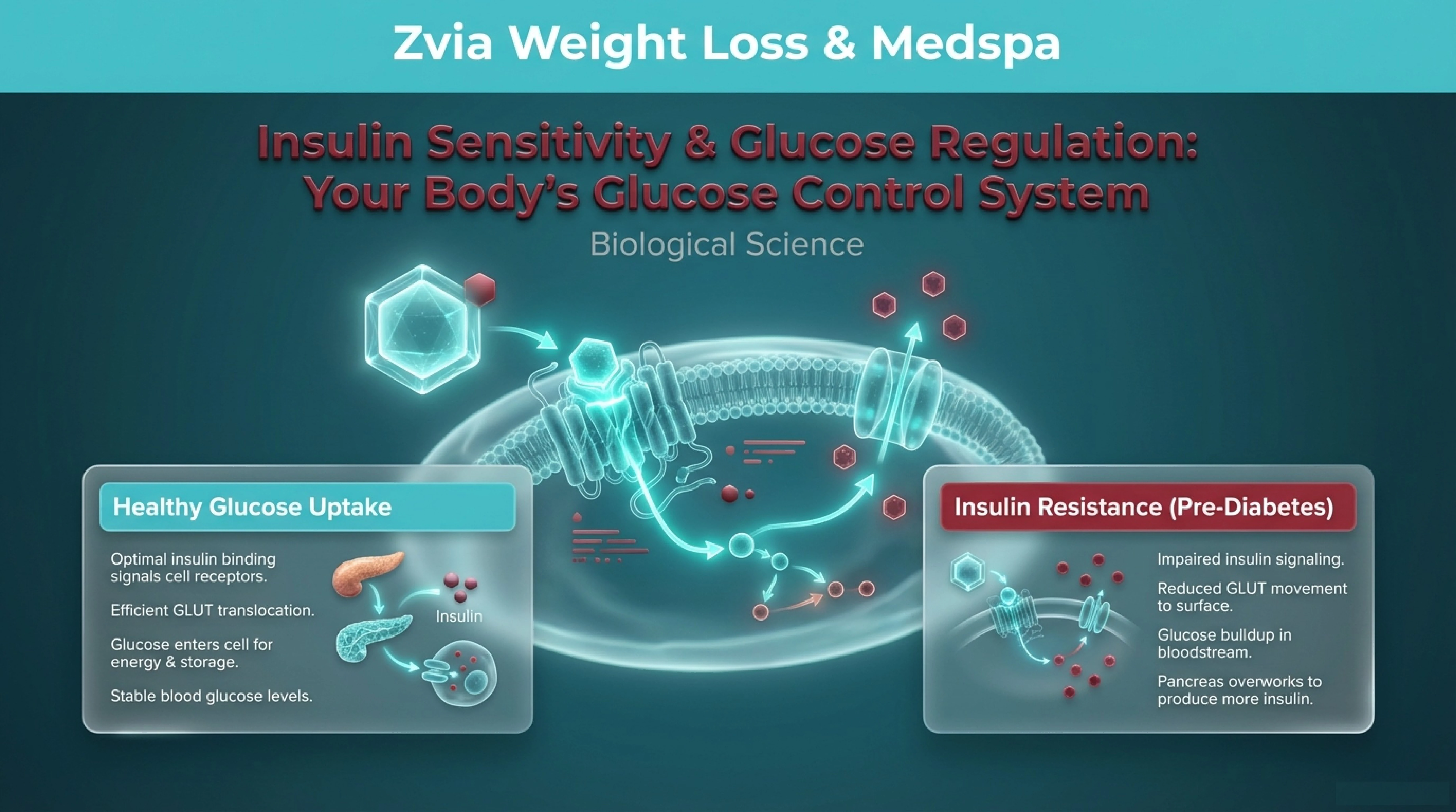
When you eat, rising blood glucose triggers your pancreatic beta cells to release insulin. Insulin travels to its three primary target tissues — skeletal muscle, liver, and adipose tissue — where it initiates a precise molecular cascade. Insulin binds to the insulin receptor, a transmembrane receptor tyrosine kinase, which activates insulin receptor substrate proteins (IRS-1 and IRS-2). These recruit and activate PI3K (phosphoinositide 3-kinase), which generates PIP3 at the cell membrane [S1]. PIP3 serves as a docking site for protein kinase B (Akt), which is then activated through dual phosphorylation by PDK1 and mTORC2 [S2].
Activated Akt phosphorylates a protein called AS160 (TBC1D4), which releases Rab GTPases from their inactive state. This triggers GLUT4 glucose transporters — stored in intracellular vesicles — to migrate to the cell surface, where they facilitate glucose entry into the cell [S1, S2]. This entire cascade, from receptor binding to glucose entry, is the molecular definition of insulin sensitivity. When it works efficiently, small amounts of insulin produce robust glucose clearance. When any step falters, the body compensates by producing more insulin — the hallmark of insulin resistance.
The Pancreatic Control Center
Glucose homeostasis is maintained by three cell types in the pancreatic islets of Langerhans working in concert. Beta cells release insulin when glucose rises. Alpha cells release glucagon when glucose falls, signaling the liver to produce glucose through glycogenolysis (glycogen breakdown) and gluconeogenesis (new glucose synthesis from precursors like lactate and amino acids) [S7, S8]. Delta cells release somatostatin, which inhibits both insulin and glucagon, serving as a regulatory brake.
These cells engage in a paradoxical paracrine feedback system: beta cell products (insulin, zinc, GABA) inhibit alpha cells, while glucagon from alpha cells stimulates beta cells [S7]. This asymmetric feedback enables the system to maintain remarkably stable blood glucose and dampen overshoots after meals.
The Liver as Metabolic Switchboard
Your liver is responsible for approximately 90% of endogenous glucose production [S19]. During fasting, gluconeogenesis contributes an estimated 54% of hepatic glucose output after 14 hours, rising to 84% after 42 hours [S19]. After a meal, insulin suppresses hepatic glucose production through a pathway converging on the transcription factor FOXO1 — when Akt phosphorylates FOXO1, it is exported from the nucleus, silencing gluconeogenic genes and shutting down unnecessary glucose production [S20].
The Incretin Amplifier
Your gut plays a surprisingly central role. After eating, intestinal L-cells release GLP-1 and K-cells release GIP — incretin hormones that account for an estimated 50-70% of total postprandial insulin secretion [S21, S22]. GLP-1 also suppresses glucagon, slows gastric emptying, and promotes satiety through vagal nerve pathways connecting the gut to brainstem appetite centers [S21]. This gut-pancreas-brain communication axis is fundamental to how your body coordinates its response to food.
The Exercise Bypass
One of the most significant discoveries in metabolic biology is that skeletal muscle possesses an insulin-independent pathway for glucose uptake during exercise. Muscle contraction activates AMPK (AMP-activated protein kinase), which drives GLUT4 translocation through a completely separate signaling route from insulin [S14, S15]. Additional contraction-mediated pathways through calcium/CaMKII and the GTPase Rac1 provide built-in redundancy [S16]. Critically, this contraction pathway remains fully functional even when insulin signaling is impaired — GLUT4 translocation during exercise is normal in individuals with insulin resistance [S14]. This is why physical activity is uniquely powerful as a metabolic intervention.
Key Benefits of Understanding This Biology
Energy Stability Has a Molecular Explanation
When insulin sensitivity is optimized, glucose enters cells efficiently, providing steady fuel. When sensitivity declines, glucose fluctuations produce the energy crashes, brain fog, and mid-afternoon fatigue that so many people experience — and often attribute to willpower rather than biology.
Body Composition Responds to Metabolic Signals
Insulin is a master regulator of fat metabolism. Chronically elevated insulin — the body’s compensation for declining sensitivity — actively promotes fat storage and suppresses fat breakdown. Understanding this mechanism explains why calorie restriction alone often fails to produce the body composition changes people expect.
The Post-Exercise Window Is Metabolically Significant
After exercise, the signaling proteins TBC1D1 and TBC1D4 remain phosphorylated for hours, priming your cells for enhanced insulin-stimulated glucose uptake [S14]. This molecular priming means insulin sensitivity can remain elevated for up to 16 hours following a single exercise session — a powerful biological tool for metabolic optimization.
Your Circadian Clock Shapes Metabolic Response
Human adipose tissue demonstrates peak insulin sensitivity around midday — approximately 54% higher than at midnight [S13]. This circadian rhythm, coordinated by your central clock and peripheral tissue clocks, means the same meal produces a meaningfully different metabolic response depending on when you eat it.
Sleep Is a Metabolic Intervention
Sleep restriction reduces insulin-stimulated Akt phosphorylation by approximately 30% [S12] — comparable to the metabolic impact of significant weight gain. When sleep loss includes circadian misalignment (shifted timing), the reduction is nearly twice as large [S12].
What to Expect: How This Biology Unfolds Across Life
The Baseline: Your 20s and Early 30s
In a metabolically healthy state, this system operates with elegant efficiency. Meals produce modest glucose rises, proportionate insulin responses, and rapid clearance. Energy is stable. Body composition responds predictably to diet and activity. The circadian metabolic rhythm runs strong.
The Shift: Your Late 30s Through 50s
Muscle mass declines approximately 3-5% per decade without targeted resistance training, reducing the body’s primary glucose disposal tissue. For women, perimenopause introduces estrogen fluctuations that directly disrupt insulin signaling — estrogen enhances insulin sensitivity through ERalpha-PI3K-Akt signaling [S25], and its decline accelerates resistance independently of age [S26]. Visceral fat redistribution, cortisol elevation, and beta cell demand all compound these changes. This is typically when people first notice that their body is “not responding the same way.”
The Adaptation: Your 50s and Beyond
The body enters a compensatory phase — beta cells increase insulin output to maintain normal glucose, a state called compensatory hyperinsulinemia. This can maintain normal blood sugar for years while silently promoting fat storage, suppressing fat breakdown, and perpetuating the conditions that drive further resistance. Understanding this trajectory is empowering because every lifestyle lever described below remains actionable.
Who Should Understand This?
If you have noticed that your energy does not sustain the way it used to, that weight has shifted in ways that feel disconnected from your habits, or that your body’s response to food and exercise has changed — this biology is likely part of the story. Specifically:
Adults navigating midlife body composition changes — particularly around the midsection — often find that understanding insulin’s role as a fat-storage regulator reframes the problem entirely. It is rarely about eating less or moving more. It is about how efficiently your cells are processing the fuel you give them.
Women in perimenopause and menopause face a convergence of estrogen decline, progesterone fluctuation, and cortisol elevation that collectively accelerate insulin resistance [S25, S26]. Understanding why these hormonal transitions reshape metabolism helps distinguish between what is modifiable and what requires clinical evaluation.
Shift workers and people with irregular sleep schedules face chronically elevated insulin resistance risk due to circadian misalignment [S12]. The research demonstrates that timing effects are distinct from and additive to sleep loss effects.
Active adults over 40 benefit from understanding why combined training — aerobic plus resistance — produces metabolic outcomes that are more than additive [S18], and why the exercise-AMPK pathway provides a glucose disposal route that remains fully functional regardless of insulin sensitivity status [S14].
Your Zvia provider can evaluate your specific metabolic markers and help determine how these biological systems are expressing in your body right now.
Working With This Biology
Nutrition as Metabolic Signaling
Meal timing aligns with circadian biology. Earlier meals correspond with your body’s peak insulin sensitivity window. Cross-sectional and experimental research consistently demonstrates lower postprandial glucose responses when the same meal is consumed earlier in the day versus evening [S23].
Dietary fiber serves multiple metabolic roles. Soluble fiber slows glucose absorption, blunting postprandial spikes. Fermentable fibers produce short-chain fatty acids — particularly butyrate — that support gut barrier integrity, reduce systemic inflammation, and modulate incretin hormone secretion [S28]. Omega-3 fatty acids activate AMPK in skeletal muscle and adipose tissue, improve mitochondrial function, and reduce endoplasmic reticulum stress [S27]. Magnesium participates in insulin receptor signaling, GLUT4 trafficking, and over 300 enzymatic processes relevant to glucose metabolism.
The Mediterranean dietary pattern demonstrates robust clinical evidence. The PREDIMED trial (722 participants) showed fasting glucose reductions of 0.39 mmol/L with olive oil supplementation, while the ATTICA study found a 27% improvement in the HOMA insulin resistance index with higher dietary adherence [S24].
Movement as Metabolic Medicine
Aerobic exercise activates the AMPK-PGC1alpha pathway, driving mitochondrial biogenesis, increased GLUT4 expression, and enhanced oxidative capacity. Resistance training increases the body’s total insulin-sensitive tissue mass. The STRRIDE trial demonstrated that combined training produced the largest improvements in insulin sensitivity, body composition, and metabolic syndrome markers compared to either modality alone [S18]. Even seven days of vigorous exercise produced significant insulin sensitivity improvements in individuals with established resistance [S17].
Sleep as Metabolic Infrastructure
Experimental studies consistently show that sleep restriction reduces insulin-stimulated glucose uptake by approximately 30% [S12]. The adipose tissue circadian rhythm — with amplitude correlated to sleep duration and inversely to late bedtimes [S13] — makes sleep duration and timing a direct lever on metabolic function.
Stress Management as Metabolic Strategy
Cortisol directly stimulates hepatic gluconeogenesis while reducing glucose uptake in muscle and adipose tissue. Chronic HPA axis activation creates sustained metabolic disruption — elevated glucose, increased visceral fat deposition, and amplified inflammatory signaling that further impairs insulin sensitivity. During hormonal transitions, declining progesterone reduces cortisol buffering, amplifying these effects.
The Zvia Perspective
At Zvia Weight Loss & MedSpa in Lakewood, Colorado, understanding insulin sensitivity and glucose regulation is not academic — it is foundational to how we approach metabolic health, body composition, and the full spectrum of health goals our clients bring to us.
We see this biology every day in our practice. The client whose weight shifted at 42 and will not respond to the strategies that worked at 32. The woman in perimenopause whose energy, body composition, and glucose patterns are all changing simultaneously. The active professional whose performance plateaued despite doing everything “right.”
These are not character failures. They are biological realities — and they have biological explanations rooted in the pathways, feedback loops, and hormonal interactions described above.
This is why Zvia invests in biological education. Not to promote a specific intervention, but because understanding the science is the first step toward meaningful outcomes. The second step is working with a team that reads your labs, interprets your metabolic markers, and builds a strategy around your unique biology — not a template.
At Zvia Weight Loss & MedSpa, metabolic health is never one-size-fits-all. Every body has its own insulin sensitivity profile, its own circadian rhythm, its own hormonal landscape. Our providers evaluate these factors with clinical precision because understanding your specific biology is the only way to design a strategy that actually works for you.
Continue exploring
Protein Synthesis & Lean Mass: Why Your Body Loses Muscle During Weight Loss
Why your body loses muscle during weight loss — and the biology of preserving lean mass. Provider-guided strategies at Zvia Weight Loss & MedSpa in Lakewood, CO.
Satiety Signaling: Why Your Hunger Stopped Making Sense
The science of satiety signaling — hormones, nerves, and brain circuits that govern hunger and fullness. Learn how your appetite biology works at Zvia Weight Loss & MedSpa in Lakewood, CO.
Educational purposes only. Provider-supervised protocols required. Results may vary based on individual biological response.
Talk with our clinical team about how this applies to your health picture.